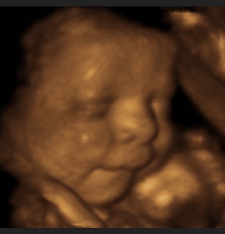
What is a 4D scan?
The 4D baby scan can be performed any time from 12 weeks but ideally between 26 to 32 weeks of pregnancy. Our team puts together the 3D images into 4D for parents-to-be to see their little one in real-time. Simply, this is a kind of ultrasound imaging, through which the baby can be seen while moving. Remember that in creating a 4D image, the 3D images are blended to create the 4D real-time scan. With 3D and 4D scans, you see your baby's skin but not the baby’s insides. You may see the shape of your baby's mouth and nose, or be able to spot the baby yawning or sticking their tongue out.
When can I have a 3D/4D scan?
This scan could be performed between 26 to 32 weeks of pregnancy. This medical imaging will be able to see his/her face, eyes, mouth and nose, and watch him/her stretch, yawn, or even suck a thumb! The further along you get in your pregnancy, the harder it may be to see the baby's face. For twins, 26 weeks is the ideal time to see the little ones. Seeing the baby's face is more difficult if the baby has arms or legs in front of it.
What is a 2D Ultrasound?
The routine 2D obstetric ultrasound creates a black and white image that shows the skeletal structure of the baby and makes the internal organs visible. As the most common form of ultrasound, a 2D ultrasound can be done during any trimester but is often performed early in gestation to confirm the pregnancy and to help establish the due date. The 2D ultrasound is most commonly used to diagnose the health of the baby. Often, the 2D scan is repeated at 18-20 weeks of gestation to check for normal growth and development
As the name implies, all 2D images are flat and have no depth to them.
It is important to remember that all images are dependent on the baby's position, movement, size, and the amount of surrounding fluid.
Is a 3D/4D ultrasound safe for me and my baby?
3D/4D ultrasound scans are always safe for both the mother and the baby.3D and 4D scans are considered as safe as 2D scans because the images are made up of sections of two-dimensional images converted into a picture. They are performed using high-quality, advanced ultrasound equipment.
How long does the 3D/4D ultrasound take?
For a good view of the baby's face, the scan may take a little longer than 40 minutes. If the baby is not in a great position, you might be asked to take a walk and return later, to have a drink, or to eat food to encourage some extra movement from the baby.
What if we are unable to see the baby’s face?
After 32 weeks or so your baby's head may go deep down in your pelvis, so you may not be able to see his/her face. Also, if the baby is facing your back, her head's far down in your pelvis, or there's not much fluid around her, you won't see much. This is also true if you have a lot of abdominal fat.
Some of our private partners may offer you a free repeat scan if you can’t see your baby’s face. However, others will advise you of the limits of ultrasound and may not offer to repeat the scan.
More than one baby?
If you're pregnant with twins or more, it's best to have a 3D or 4D scan earlier rather than later, when you're about 27 weeks pregnant, for a session to allow enough room for both babies to be seen. This should allow you to get a clearer view of each baby.
If the baby is lying facing outwards, you should be able to see the face clearly. But if she's facing your back, or if the head is far down in your pelvis, you may not get a clear view of the baby. This is also true if there is a lot of belly fat
The sonographer may ask you to go for a walk, or to come back in a week, when your baby may have moved to a better position.
Early Gender Sneak Peak
Many of our locations offer early gender determination which is one of our specialties. Prenatal Sneak Peek, 3D 4D Ultrasounds provide elective 3D ultrasounds to meet your precious unborn baby and get a preview of your little one’s face, in a 3D world. A Gender Determination session may be completed once you have reached the 14-week mark. Is it a boy or girl? Who does the baby look like?
Other findings:
If our staff finds anything abnormal, those will be discussed with the physician who will then discuss the case with you, ensuring further investigation and intervention as necessary.
